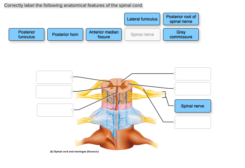
44 correctly label the anatomical features of the spinal cord
anatomy of the human spinal cord Quiz - PurposeGames.com This is an online quiz called anatomy of the human spinal cord, There is a printable worksheet available for download here so you can take the quiz with pen and paper. This quiz has tags. Click on the tags below to find other quizzes on the same subject. Anatomy, labeling, Your Skills & Rank, Total Points, 0, Get started! Today's Rank, --, 0, Correctly label the following anatomical features of the eye. Correctly ... Correctly Label The Following Anatomical Features Of The Spinal Cord. Lateral Funiculus Posterior Root Of Spinal Nerve Posterior Funiculus Posterior Horn Anterior Median Fissure Spinal Nerve Gray Commissure Spinal Nerve (B) Spinal Cord And...
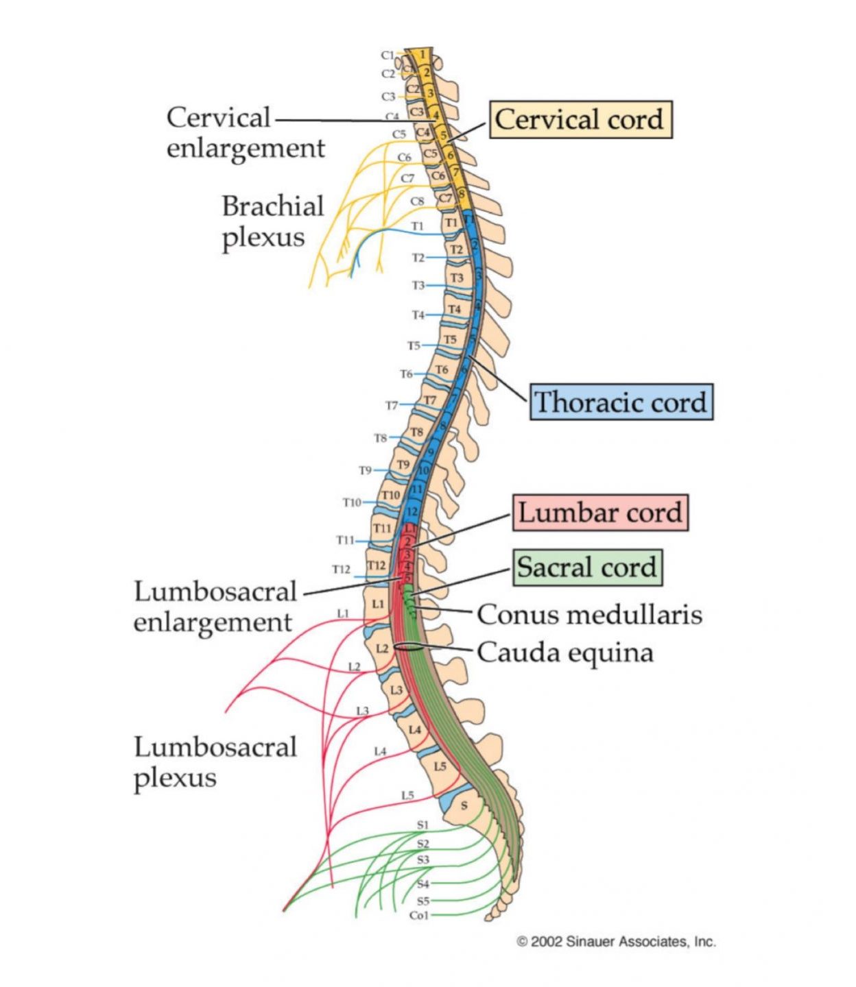
Anatomy, Central Nervous System - StatPearls - NCBI Bookshelf The spinal cord ends in a cone-shaped structure called conus medullaris and is supported to the end of the coccyx by the filum terminale. Ligaments are found throughout the spinal column, securing the spinal cord from top to bottom. Ascending pathway to the brain: Sensory information travels from the body to the spinal cord before reaching the ...

Correctly label the anatomical features of the spinal cord
An automated treatment planning framework for spinal radiotherapy and ... Using each contour pair, the following 9 features were calculated: differences in contour dimension (dx, dy, dz), surface DSC (from Nikolov et al 25 ), DSC, 95 th percentile Hausdorff distance (HD95), adjacent vertebral body contour overlap of the primary contour, rate of change of vertebral spacing of the primary contour (defined in XXXX et al ... Neural pathways and spinal cord tracts: Anatomy | Kenhub The gracilis and cuneate fasciculi, also known as the dorsal/posterior columns, are two ascending pathways located side-by-side in the posterior funiculus of the spinal cord. They carry fine and discriminative touch as well as proprioceptive sensations. Correctly label the following anatomical features of the spinal cord. Correctly label the following anatomical features of the spinal cord. posted on February 26, 2022
Correctly label the anatomical features of the spinal cord. Spinal Cord Cross Section | New Health Advisor 2-Minute Neuroscience: Spinal Cord Cross-section, 1. White Matter, The white matter has the nerve fibers that run up and down the length of the cord, they are called axons. This makes it possible for the different parts of the CNS communicate with each other. Every bundle of axons is a tract and it transmits specific information. Procedure Bstructure Of The Spinal Cord - Human Anatomy - GUWS Medical 1. Review a textbook section on the spinal cord. 2. As a review activity, label figures 27.1, 27.2, and 27.3. 3. Complete Parts B and C of the laboratory report. 4. Obtain a prepared microscope slide of a spinal cord cross section. Use the low power of the microscope to locate the following features: Spinal Cord Injury Levels: A Complete Overview of Each Type - Flint Rehab C7 spinal cord injury - increasing sensation in the hand, including intact sensation of the middle finger. Those with a C7 SCI will be able to straighten the elbows and bend the wrists. C8 spinal cord injury - will be able to bend the fingers and grasp objects. Sensation and most movement in the hand will be intact at this level of injury. Spinal nerves: Anatomy, roots and function | Kenhub They are composed of both motor and sensory fibres, as well as autonomic fibres, and exist as 31 pairs of nerves emerging intermittently from the spinal cord to exit the vertebral canal. This article will discuss the anatomy and function of the spinal nerves. Contents, Terminology, Anterior (ventral) and posterior (dorsal) roots,
Spinal Reflex: Anatomy and Examples | Kenhub The first is located within the spinal ganglion. This is the sensory neuron (afferent) whose peripheral process detects the stimuli from the muscle. Then, the central process of the first neuron conducts this signal to the ventral horn of the spinal cord, where the second neuron is situated. Spinal cord: Ascending and descending tracts | Kenhub The spinal cord is a cylindrical mass of neural tissue extending from the caudal aspect of the medulla oblongata of the brainstem to the level of the first lumbar vertebra (L1). While the length of the spinal cord varies from one individual to another, it is usually longer in males (approximately 45 cm) than it is in females (approximately 42 cm). Central nervous system: Anatomy, structure, function | Kenhub They are enveloped and protected by three layers of meninges, and encased within two bony structures; the skull and vertebral column, respectively. The brain consists of the cerebrum, subcortical structures, brainstem and cerebellum. The spinal cord continues inferiorly from the brainstem and extends through the vertebral canal. Anatomy of the spinal cord - e-Anatomy - IMAIOS The first image shows the different segments of the spinal cord (cervical, thoracic, lumbar, sacral and coccygeal segments), the emergence of spinal nerves (cervical, thoracic, lumbar and sacral nerves and coccyx at the level of the cauda equina and filum terminale) and the sectional aspect of the spinal cord with changes in diameter at the cerv...
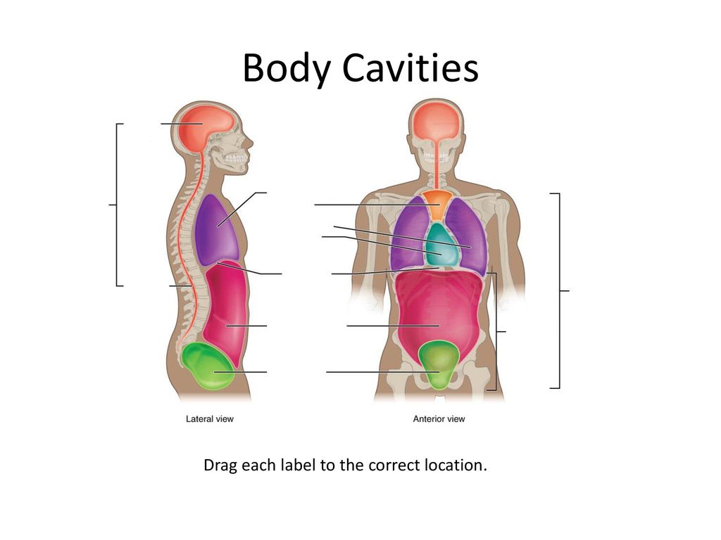
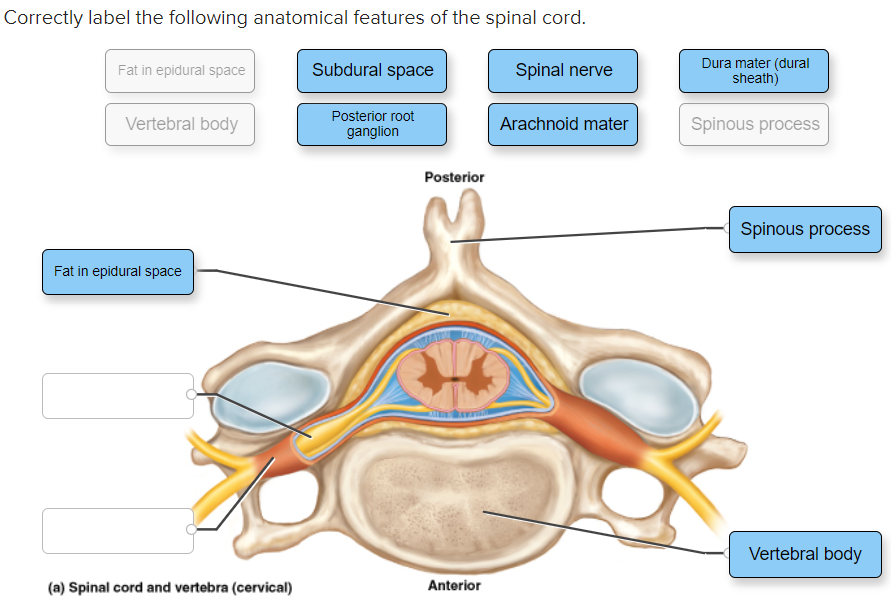
Body Cavities and Membranes: Labeled Diagram, Definitions - EZmed Spinal Cavity, Thoracic Cavity, Pleural Cavities, Pericardial Cavity, Abdominopelvic Cavity, Abdominal Cavity, Pelvic Cavity, You will learn the main organs housed in each cavity, along with the membranes that line the cavity and the type of fluid present. Labeled diagrams, lateral views, and concise explanations included! Correctly label the following anatomical features of the spinal cord ... Correctly label the following anatomical features of the spinal cord. Fat in epidural space Subdural space Spinal nerve Dura mater (dural sheath) Vertebral body Posterior root ganglion Arachnoid mater Spinous process Posterior Spinous process Fat in epidural space Vertebral body (a) Spinal cord and vertebra (cervical) Anterior, Spinal Nerves: Anatomy, Function, and Treatment - Verywell Health The spine is made up of vertebrae (back bones) that protect and surround the spinal cord, which is a column of nerve tissue. Spinal nerves branch out from the spinal cord. These are peripheral nerves, or those that run through other parts of the body and transmit message to and from the brain/spinal cord. Spine anatomy diagrams and interactive vertebrae quizzes | Kenhub The vertebral column (spine) extends from the inferior aspect of the occipital bone of the skull to the tip of the coccyx. It can be divided into 5 regions, each characterized by different types of vertebrae: Cervical region (7 vertebrae) Thoracic region (12 vertebrae) Lumbar region (5 vertebrae) Sacral region (5 fused vertebrae)
Spinal cord: Anatomy, structure, tracts and function | Kenhub The spinal cord is made of gray and white matter just like other parts of the CNS. It shows four surfaces: anterior, posterior, and two lateral. They feature fissures (anterior) and sulci (anterolateral, posterolateral, and posterior). The gray matter is the butterfly-shaped central part of the spinal cord and is comprised of neuronal cell bodies.
A harmonized atlas of mouse spinal cord cell types and their ... - Nature a Summary of the datasets used in this study, including the studies that used single-cell/nucleus RNA sequencing to analyze postnatal mouse spinal cord cell types (colored names above the gray bar ...
Correctly label the following anatomical features of the spinal cord. Correctly label the following anatomical features of the spinal cord. posted on February 26, 2022
Neural pathways and spinal cord tracts: Anatomy | Kenhub The gracilis and cuneate fasciculi, also known as the dorsal/posterior columns, are two ascending pathways located side-by-side in the posterior funiculus of the spinal cord. They carry fine and discriminative touch as well as proprioceptive sensations.
An automated treatment planning framework for spinal radiotherapy and ... Using each contour pair, the following 9 features were calculated: differences in contour dimension (dx, dy, dz), surface DSC (from Nikolov et al 25 ), DSC, 95 th percentile Hausdorff distance (HD95), adjacent vertebral body contour overlap of the primary contour, rate of change of vertebral spacing of the primary contour (defined in XXXX et al ...













:watermark(/images/watermark_5000_10percent.png,0,0,0):watermark(/images/logo_url.png,-10,-10,0):format(jpeg)/images/overview_image/1900/4wg7EtKVwtWY7wcLa4OtAA_anatomy-spinal-cord-cross-section_english.jpg)




Komentar
Posting Komentar